Beyond Visual Inspection: Using Micro-XCT and AI for Flawless Impregnation Efficacy Assessment
This technical summary is based on the academic paper "Micro X-ray Computed Tomography and Machine Learning Assessment of Impregnation Efficacy of Die-Casting Defects in Metal Alloys" by Ajith Bandara, Koichi Kan, Katanaga Yusuke, Natsuto Soga, Takagi Katsuyuki, Akifumi Koike, and Toru Aoki, published in Sensors and Materials (2024). It has been analyzed and summarized for technical experts by CASTMAN.


Keywords
- Primary Keyword: Impregnation Efficacy Assessment
- Secondary Keywords: Micro X-ray CT, Machine Learning Segmentation, Die-Casting Defects, Vacuum Pressure Impregnation (VPI), ADC12 Alloy, Nondestructive Testing (NDT)
Executive Summary
- The Challenge: Reliably verifying that Vacuum Pressure Impregnation (VPI) has successfully sealed microscopic leakage paths and casting defects in die-cast components is a critical and difficult challenge for ensuring product airtightness.
- The Method: Researchers used advanced micro X-ray computed tomography (micro-XCT) with direct conversion CdTe sensors and a machine learning (ML) segmentation model to non-destructively visualize and analyze impregnated Al-alloy samples.
- The Key Breakthrough: The machine learning approach successfully segmented and identified sealant resin within subtle leakage paths (20-50 µm) that are nearly invisible and difficult to distinguish using traditional intensity-based CT image analysis.
- The Bottom Line: Micro-XCT combined with machine learning provides a powerful, quantitative, and non-destructive method to precisely validate the Impregnation Efficacy Assessment, ensuring component reliability and improving quality assurance.
The Challenge: Why This Research Matters for HPDC Professionals
In the high-stakes world of HPDC for automotive, aerospace, and other critical industries, airtightness is not a luxury—it's a necessity. Defects like gas pores and shrinkage cracks can create interconnected leakage paths, leading to component failure. While Vacuum Pressure Impregnation (VPI) is a cost-effective method for sealing these defects, verifying its success is a significant challenge. Traditional methods like the dipping water leak test are often inadequate for ensuring complete impregnation, especially in complex, microscopic pathways.
The core problem is one of visibility. The polymer resin used for sealing has a low atomic number and density, making it difficult to detect with conventional X-ray techniques. Its low grey contrast in CT images makes it hard to distinguish from air pockets, leading to uncertainty about the quality of the seal. This research was undertaken to develop a reliable, non-destructive imaging technique to overcome these limitations and provide a clear, in-depth analysis of impregnation efficacy.
The Approach: Unpacking the Methodology
The researchers conducted their investigation on three cuboidal ADC12 aluminum alloy samples (10 × 10 × 5 mm³), which were impregnated using a standard VPI technique (MIL-STD-276A Method B) with a commercial thermosetting polymer, Super Seal P601.
The primary analytical method was a laboratory-based micro-XCT system featuring two types of advanced cadmium telluride (CdTe) sensors:
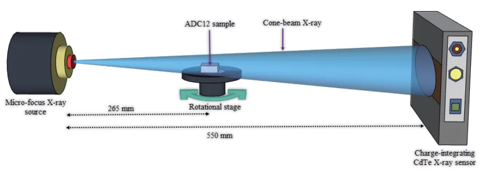
1. Qualitative Analysis: A charge-integration-type CdTe sensor was used to capture 2700 projection images per sample. These images were reconstructed into 3D and 2D CT scans. A machine learning tool, Trainable Weka Segmentation (TWS), was then applied to precisely segment the Al alloy, air pores, and sealant resin, overcoming the limitations of traditional intensity-based analysis.
2. Quantitative Analysis: A photon-counting CdTe sensor was used for dual-energy XCT (DXCT). By capturing data in low-energy (20–30 keV) and high-energy (50–60 keV) windows, the researchers could calculate the effective atomic number (EAN) of the impregnation material, allowing for its positive identification.
This dual approach provided both a detailed visualization of the sealant distribution and a quantitative confirmation of the material itself.
The Breakthrough: Key Findings & Data
The study yielded several crucial findings that provide a new level of insight into the VPI process.
Finding 1: Machine Learning Overcomes the Limits of Conventional CT Imaging
Traditional CT image analysis struggles to separate the low-density sealant from air and the Al alloy. The study quantified this challenge, calculating a low image quality measure (Q) of just 6.26 between air and sealant (Table 1), indicating poor separation. However, the machine learning-based TWS approach successfully overcame this. As shown in Figure 7, the ML model clearly segmented a subtle, successfully sealed internal leakage path of just 20-50 µm in sample 2. The original CT image (Fig. 7b) shows a nearly invisible path, while the segmented image (Fig. 7c) clearly delineates the alloy (red), sealant (green), and pores (purple), providing unambiguous proof of a successful seal.
Finding 2: Visualization of Incomplete Impregnation and Process Defects
The analysis of sample 3 highlighted the importance of this advanced Impregnation Efficacy Assessment. The 3D and 2D CT images (Figure 8) revealed a significant die-casting crack with a discontinuous seal. The sealant resin was confined to only a narrow region (40-130 µm) of the leakage path. The study suggests that the low-viscosity resin may have drained from the larger defect during the standard VPI process. Furthermore, the magnified ML-segmented image (Figure 9a) revealed minute air pockets trapped within the impregnated sealant, which could be caused by polymerization shrinkage or moisture and can compromise the seal's integrity. This finding demonstrates that even when a VPI process is performed, it may not be 100% effective in all cases.
Finding 3: Quantitative Identification of Sealant Material
Using the DXCT method, the researchers quantitatively identified the sealant material within the sample. By analyzing the linear attenuation coefficients in different energy windows, they calculated the experimental effective atomic number (EAN) of the resin. As detailed in Table 2, the experimental EAN (Zeff(exp)) was 7.36. This value closely matched the theoretical EAN (Zeff(th)) of 7.10 for the P601 Super Sealant, with a minimal error margin of 3.53%. This result provides quantitative validation, confirming that the material observed in the defects is indeed the intended sealant.
Practical Implications for R&D and Operations
- For Process Engineers: This study suggests that standard VPI process parameters may not be universally effective for all defect sizes. The observation of sealant draining from a larger crack in sample 3 (Figure 8) indicates that adjusting process parameters like pressure cycles or sealant viscosity may be necessary to improve sealing efficacy for certain types of defects.
- For Quality Control Teams: The data in Figure 7 and Table 1 of the paper illustrates that this ML-enhanced micro-XCT method can non-destructively verify sealing in defects as small as 20 µm, far beyond the capability of standard leak tests. This could inform new, more robust quality inspection criteria for critical components where 100% airtightness is mandatory.
- For Design Engineers: The findings indicate that the geometry of casting defects significantly influences impregnation success. The visualization of the complex, irregular crack in sample 3 (Figure 8) suggests that design features prone to creating such defects should be carefully reviewed, as they may be difficult to seal effectively.
Paper Details
Micro X-ray Computed Tomography and Machine Learning Assessment of Impregnation Efficacy of Die-Casting Defects in Metal Alloys
1. Overview:
- Title: Micro X-ray Computed Tomography and Machine Learning Assessment of Impregnation Efficacy of Die-Casting Defects in Metal Alloys
- Author: Ajith Bandara, Koichi Kan, Katanaga Yusuke, Natsuto Soga, Takagi Katsuyuki, Akifumi Koike, and Toru Aoki
- Year of publication: 2024
- Journal/academic society of publication: Sensors and Materials, Vol. 36, No. 1
- Keywords: micro X-ray computed tomography, direct conversion X-ray sensors, machine learning image segmentation, Al-alloy die-casting, vacuum pressure impregnation, dual-energy X-ray CT
2. Abstract:
Die-cast light metal alloys in various industrial applications require precise airtightness, and vacuum pressure impregnation (VPI) is typically used to seal casting defects to ensure product reliability. Evaluating the efficacy of VPI in sealing alloy defects is crucial. In this study, laboratory-based micro X-ray computed tomography (micro-XCT) was effectively employed in conjunction with advanced direct conversion CdTe semiconductor sensors to nondestructively evaluate the efficacy of standard VPI in sealing die-casting defects of industrial Al alloys. The internal casting defects and the low-atomic-number impregnation sealant distribution were visualized by adjusting the scalar opacity mapping in 3D CT. In 2D CT, it is challenging to identify the sealant resin in the narrow leakage paths of the alloy sample due to its low grey contrast, and a machine learning approach with the Trainable Weka Segmentation (TWS) plug-in was applied to segment the CT images more precisely than by the traditional intensity-based image processing technique. TWS efficiently segmented the Al alloy, air pores, and diffused sealant resin in the samples, providing an in-depth analysis of the impregnation efficacy. Dual-energy XCT (DXCT) with photon-counting sensors was utilized as a quantitative method based on the effective atomic number to identify the impregnation material in the alloys as the commercially used Super Sealant P601 polymer resin.
3. Introduction:
X-ray computed tomography (XCT) has become a broad and effective nondestructive imaging technique used in the medical and industrial sectors since the first commercial CT scanner was built for medical imaging by Nobel Prize winner Godfrey Hounsfield in 1969. With the rapid development of CT technology, computer performance, and software, XCT in the industrial field has become well known for its faster and more cost-effective investigation than traditional evaluation methods. As a robust nondestructive testing (NDT) method, XCT is increasingly widely used in various industrial sectors, particularly the automotive, aerospace, and material industries. The authors have been investigating the efficacy of the impregnation technique in sealing die-casting defects of light metal alloys. Such die-cast alloys are widely used for intricate industrial parts, especially in automotive, owing to advantages including reduced weight, enhanced fuel efficiency, corrosion resistance, high strength-to-weight ratio, thermal conductivity, and recyclability. However, high-pressure die-casting (HPDC) generates scraps due to defects such as gas pores and shrinkage cracks. Interconnected pores and leakage paths often result in the leakage of gases, liquids, and oil. Despite advances in die-casting technology, completely eliminating defects remains a significant challenge. Hence, impregnation treatments are applied as a cost-effective means of sealing pores and eliminating leakage paths. Nevertheless, the current standard dipping water leak test is inadequate to ensure impregnation efficacy, and a reliable, nondestructive imaging technique is required.
4. Summary of the study:
Background of the research topic:
Die-cast light metal alloys require precise airtightness for many industrial applications. Vacuum pressure impregnation (VPI) is the standard method used to seal casting defects, but evaluating its effectiveness is crucial and challenging.
Status of previous research:
While micro-XCT is widely used for analyzing structures and imperfections in metal alloys, its potential for visualizing and characterizing low-density impregnation resin within sealed defects has been largely unexplored. This is due to the resin's low X-ray attenuation, making it difficult to detect with adequate contrast and resolution.
Purpose of the study:
To investigate the efficacy of VPI in sealing Al alloy (ADC12) die-casting defects using a laboratory-based micro-XCT system equipped with advanced direct conversion CdTe semiconductor sensors. The study aimed to combine this with a machine learning approach for more precise image segmentation and dual-energy XCT for quantitative material identification.
Core study:
The study involved performing micro-XCT on three impregnated ADC12 samples. The researchers used 3D CT with scalar opacity mapping and 2D CT analysis to visualize defects and sealant distribution. A machine learning tool (Trainable Weka Segmentation) was employed to segment the Al alloy, air pores, and sealant resin. Finally, dual-energy XCT was used to calculate the effective atomic number (EAN) of the sealant to quantitatively identify it as the commercial Super Sealant P601.
5. Research Methodology
Research Design:
The study employed two different XCT methods for a comprehensive assessment. A qualitative analysis was conducted using a charge-integration-type pixeled CdTe X-ray sensor to visualize the sealant distribution and segment the images using machine learning. A quantitative analysis was then performed using a dual-energy, photon-counting CdTe sensor to identify the sealant material based on its EAN.
Data Collection and Analysis Methods:
For qualitative analysis, 2700 projection images were captured for each sample over a 360° rotation using a cone-beam CT (CBCT) geometry. Images were reconstructed using a filtered back projection (FBP) algorithm. Image analysis was performed using Fiji and 3D Slicer software, and segmentation was done with the Trainable Weka Segmentation (TWS) tool. For quantitative analysis, projection images were acquired at four distinct energy thresholds (above 20, 30, 50, and 60 keV) to construct low- and high-energy CT images, from which the EAN was calculated.
Research Topics and Scope:
The research focused on ADC12 Al alloy die-cast samples treated with a standard VPI process using Super Sealant P601. The scope included the non-destructive visualization of internal casting defects, the qualitative assessment of sealant distribution in micro-leakage paths using machine learning, and the quantitative identification of the sealant material using dual-energy XCT.
6. Key Results:
Key Results:
- Traditional intensity-based methods are challenged in separating the low-density sealant from air and the Al alloy, evidenced by a low image quality measure (Q) of 6.26 between air and sealant.
- A machine learning (TWS) approach effectively segmented the Al alloy, air pores, and sealant resin, enabling clear visualization of successfully sealed leakage paths as narrow as 20-50 µm.
- Micro-XCT analysis revealed an instance of incomplete impregnation in a sample with a significant die-casting crack, where the sealant was confined to a narrow region and may have drained from the larger defect.
- The ML-segmented images also identified minute air pockets trapped within the sealant, a potential cause of reduced impregnation efficacy.
- Dual-energy XCT quantitatively identified the impregnation material as P601 Super Sealant by calculating its effective atomic number (EAN) to be 7.36, closely matching the theoretical value of 7.10 with a 3.53% error.
Figure Name List:
- Fig. 1. (Color online) Images of die-cast Al alloy test samples.
- Fig. 2. (Color online) Experimental setup of the charge integration-type CdTe sensor-based FPD.
- Fig. 3. (Color online) Experimental setup of the photon-counting CdTe sensor-based XCounter FPD.
- Fig. 4. (Color online) Three-dimensional CT images of ADC sample 1: (a) greyscale CT image and (b) side-view transparent CT image.
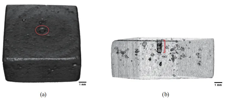
- Fig. 5. (Color online) XCT images of ADC sample 1: (a) top-view 3D CT image, (b) side-view 3D CT image, and (c) 2D CT image across the center hole.
- Fig. 6. (Color online) ML-based image segmentation of sample 1: (a) greyscale 2D CT image, (b) segmented 2D CT image, and (c) probability map of the segmented 2D CT image.
- Fig. 7. (Color online) ML-based image segmentation of sample 2: (a) 3D CT image, (b) cross-sectional 2D CT image, (c) segmented 2D CT image, and (d) probability map of the segmented 2D CT image.
- Fig. 8. (Color online) XCT images of ADC sample 3: (a) side-view 3D CT image and (b) cross-sectional 2D CT image across the highlighted region of the 3D CT image.
- Fig. 9. (Color online) ML-based image segmentation of sample 3: (a) segmented 2D CT image and (b) probability map of the segmented 2D CT image.
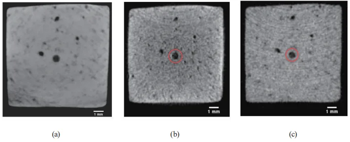
- Fig. 10. (Color online) DXCT of ADC12 sample 1: (a) total-energy 2D CT image, (b) low-energy 2D CT image, and (c) high-energy 2D CT image.


7. Conclusion:
The study successfully demonstrated that a laboratory-based micro X-ray CT system with advanced CdTe flat panel sensors is an excellent tool for nondestructively evaluating the efficacy of impregnation in sealing die-casting defects. While traditional intensity thresholding proved challenging, the ML-based image segmentation approach with TWS effectively and accurately detected the low-atomic-number sealant within subtle defects. A perfectly sealed minute leakage path (20–50 µm) was clearly depicted. The analysis also revealed an unevenly sealed leakage path in another sample, suggesting potential discharge of the sealant during the VPI process. The DXCT analysis quantitatively recognized the impregnation material as P601 Super Sealant. The outcomes confirm the effectiveness of industrial micro-XCT combined with advanced X-ray sensors to comprehensively verify impregnation efficacy.
8. References:
- [1 S. T. Neel and R. N. Yancey: Rev. Prog. Quant. Nondestr. Eval. 15 (1996) 497.]
- [2 S. Carmignato: Industrial X-ray computed tomography, W. Dewulf and R. Leach, Eds. (Springer International Publishing, Cham, Switzerland, 2017) 1st ed., Vol. 10, pp. 978-3. https://doi.org/10.1007/978-3-319-59573-3.]
- [3 S. R. Stock: Int. Mater. Rev. 53 (2008) 129. https://doi.org/10.1179/174328008X277803.]
- [4 J. G. Behnsen, K. Black, J. E. Houghton, and R. H. Worden: Materials (Basel) 16 (2023) 1259. https://doi.org/10.3390/ma16031259.]
- [5 V. Gómez, H. Herazo, and E. S. Stuart: Precis. Eng. 60 (2019) 544 https://doi.org/10.1016/j.precisioneng.2019.06.007]
- [6 F. Akman, R. Durak, M. F. Turhan, and M. R. Kaçal: Appl. Radiat. Isot. 101 (2015) 107.]
- [7 C. Cao, M. F. Toney, T.-K. Sham, R. Harder, P. R. Shearing, X. Xiao, and J. Wang: Mater. Today 34 (2020) 132.]
- [8 Y. Zhou, J. Chen, O. M. Bakr, and O. F. Mohammed: ACS Energy Lett. 6 (2021) 739.]
- [9 T. Buzug: Computed Tomography (Springer, Berlin, Germany, 2008).]
- [10 S. K. Kennedy, A. M. Dalley, and G. J. Kotyk: J. Mater. Eng. Perform. 28 (2019) 728. https://doi.org/10.1007/s11665-018-3841-5]
- [11 J. Kastner: 6th Conf. Industrial Computed Tomography 2016 (iCT2016). Case Studies in Nondestructive Testing and Evaluation, 6 (Part B) (2016) 2. https://doi.org/10.1016/j.csndt.2016.05.007.]
- [12 F. Garcia-Moreno, T. R. Neu, P. H. Kamm, and J. Banhart: Adv. Eng. Mater. 28 (2022) 201355. https://doi.org/10.1002/adem.202201355.]
- [13 D. Liu and J. Tao: Adv. Mater. Res. 308-310 (2011) 785. https://doi.org/10.4028/www.scientific.net/AMR.308-310.785.]
- [14 W. J. Joost and P. E. Krajewski: Scr. Mater. 128 (2017) 107.]
- [15 H. Juergen and T. Al-Samman: Acta Mater. 61 (2013) 818.]
- [16 F. Campbell (Ed.): Lightweight Materials: Understanding the Basics (ASM International: Novelty, OH, USA, 2012).]
- [17 J. Relland, L. Bax, and M. A. Ierdes: Vision on the Future of Automotive Lightweighting Alliance (Surrey, UK, 2019).]
- [18 F. Czerwinski: Materials 14 (2021) 6631. https://doi.org/10.3390/ma14216631]
- [19 NADCA Product Specification Standards for Die Casting, Publication #402 (Arlington Heights, IL, North American Die Casting Association, 2018) 10th ed.]
- [20 D. Blondheim and A.Monroe: Int. J. Metalcast. 16 (2022) 330. https://doi.org/10.1007/s40962-021-00602-x.]
- [21 L. Lattanzi, A. Fabrizi, A. Fortini, M. Merlin, and G. Timelli: Procedia Struct. Integrity 7 (2017) 505. https://doi.org/10.1016/j.prostr.2017.11.119]
- [22 K. Kan, Y. Imura, H. Morii, K. Kobayashi, T. Minemura, and T. Aoki: World J. Nucl. Sci. Technol. 3 (2013) 106.]
- [23 A. Bandara, K. Kan, H. Morr, A. Koike, and T. Aoki: Prod. Eng. Res. Devel. 14 (2020) 147. https://doi.org/10.1007/s11740-019-00946-8.]
- [24 N. Soga, A. Bandara, K. Kan, A. Koike, and T. Aoki: Prod. Eng. Res. Devel. 15 (2021) 885. https://doi.org/10.1007/s11740-021-01071-1.]
- [25 K. Yusuke, A. Bandara, N. Soga, K. Kan, A. Koike, and T. Aoki: Prod. Eng. Res. Devel. 17 (2023) 291. https://doi.org/10.1007/s11740-022-01147-6.]
- [26 A. D. Plessis and P. Rossouw: Case Stud. Nondestruct. Test Eval. (2015). https://doi.org/10.1016/j.csndt.2015.03.001]
- [27 A. Buratti, J. Bredemann, M. Pavan, R. Schmitt, and S. Carmignato: Applications of CT for dimensional metrology. In: S. Carmignato, W. Dewulf, and R. Leach (Eds.) Industrial X-ray computed tomography (Springer, Cham. 2018) pp. 333-369.]
- [28 J. Kastner and C. Heinzl: X-ray computed tomography for nondestructive testing and materials characterization. In: Liu Z, Ukida H, Ramuhalli P, Niel K (Eds.) Integrated imaging and vision techniques for industrial inspection (Springer, London, 2015) p. 227.]
- [29 C. Reinhart: 17th World Conf. Nondestructive Testing, Shanghai, China eJNDT 13 (2008) 25.]
- [30 A. Thompson and R. Leach: Introduction to industrial X-ray computed tomography. In: S. Carmignato, W. Dewulf, and R. Leach (Eds.) Industrial X-ray computed tomography (Springer, Cham, 2018) pp. 1-23.]
- [31 H. Randolf, T. Fuchs, and N. Uhlmann: Nucl. Instrum. Methods Phys. Res., Sect. A 591 (2008) 14.]
- [32 M. A. Krueger, S. S. Huke, and R. W. Glenny: CircRes 112 (2013) e88. https://doi.org/10.1161/CIRCRESAHA.113.301162.]
- [33 J. Maiora and M. Graña: The 2012 Int. Joint Conf. Neural Networks (IJCNN) Brisbane, Australia, June 10-15 (IEEE Explore, 2012) pp. 1–7.]
- [34 W. Macdonald and S. Shefelbine: Med. Biol. Eng. Comp. 51 (2013) 1157.]
- [35 B. Mutiargo, A. Garbout, and A. A. Malcolm: Proc. SPIE 11050, Int. Forum Medical Imaging in Asia 2019 (27 March 2019) 110500L. https://doi.org/10.1117/12.2521768]
- [36 A. Kyrieleis, V. Titarenko, M. Ibison, T. Connolley, and P. J. Withers: J. Microsc. 241 (2010) 69.]
- [37 J. Muders, J. Hesser, A. Lachner, and C. Reinhart: Int. Symp. Digital Industrial Radiology and Computed Tomography (Berlin, Germany, 2011).]
- [38 J. Schindelin, I. Arganda-Carreras, E. Frise, V. Kaynig, M. Longair, T. Pietzsch, S. Preibisch, C. Rueden, S. Saalfeld, B. Schmid, J. Y. Tinevez, D. J. White, V. Hartenstein, K. Eliceiri, P. Tomancak, and A. Cardona: Nat. Methods 9 (2012) 676. https://doi.org/10.1038/nmeth.2019]
- [39 R. Kikinis, S. D. Pieper, and K. Vosburgh: 3D slicer: a platform for subject-specifc image analysis, visualization, and clinical support. In: F. A. Jolesz (Ed.) Intraoperative imaging image-guided therapy 3 (2014) 277.]
- [40 M. Reiter, D. Weiß, C. Gusenbauer, M. Erler, C. Kuhn, S. Kasperl, and J. Kastner: Proc. 5th Conf. Industrial Computed Tomography (iCT 2014) (Wels, Austria, NDT.net) pp. 273-282.]
- [41 I. Arganda-Carreras, V. Kaynig, C. Rueden, K. W. Eliceiri, J. Schindelin, A. Cardona, and S. H. Seung: Bioinformatics 33 (2017) 2424. https://doi.org/10.1093/bioinformatics/btx180]
- [42 D. F. Jackson and D. J. Hawkes: Phys. Rep. 70 (1981) 169.]
- [43 D. J. Hawkes and D. F. Jackson: Phys. Med. Biol. 25 (1980) 1167.]
- [44 R. Nowotny: XMuDat: photon attenuation data on PC. IAEA-NDS-195 (International Atomic Energy Agency, Vienna, Austria, 1998). http://www.mds.iaea.or.at/reports/mds-195.htm.]
Expert Q&A: Your Top Questions Answered
Q1: Why was the Trainable Weka Segmentation (TWS) tool chosen over traditional intensity-based methods?
A1: Traditional intensity-based methods struggle to distinguish the low-density sealant from air and the Al alloy due to low grey contrast. The paper quantifies this with a low image quality measure (Q3) of 6.26 between air and sealant (Table 1). TWS, a machine learning approach, was trained on a known dataset from sample 1, allowing it to learn the distinct features of each material and precisely segment them even when their intensity values overlap, providing a much more accurate analysis.
Q2: The paper mentions an incomplete seal in sample 3. What was the suspected cause?
A2: The paper suggests two main possibilities for the incomplete seal shown in Figure 8. First, the low-viscosity Super Sealant resin might have drained from the comparatively significant defects during the standard impregnation technique. Second, as revealed in the magnified image in Figure 9(a), the entrapment of minute air pockets within the sealant itself, possibly due to polymerization shrinkage or moisture, could also contribute to a compromised seal.
Q3: How did the researchers quantitatively confirm the sealant material was P601?
A3: They used a technique called Dual-Energy XCT (DXCT) with a photon-counting sensor to calculate the material's effective atomic number (EAN). As shown in Table 2, the experimentally derived EAN for the sealant was 7.36. This value provided quantitative validation, as it closely matched the theoretical EAN of 7.10 for the P601 sealant with only a 3.53% error margin.
Q4: What was the size of the smallest leakage path successfully identified and visualized by the ML model?
A4: As depicted in Figure 7(b), the study successfully visualized a sealed subtle internal leakage path in Al alloy sample 2 with a width of 20-50 µm. The TWS ML-based image segmentation, shown in Figure 7(c), was crucial for clearly segmenting the sealant within this microscopic path, demonstrating the high precision of the technique.
Q5: What is the significance of the advanced CdTe semiconductor sensors used in this study?
A5: The paper highlights that the advanced direct conversion CdTe sensors were critical to the study's success. They offer several distinct advantages over conventional sensors, including higher X-ray conversion efficiency, energy differentiation via photon counting (which enables DXCT), and diffusion-free imaging. These features facilitate the creation of the high-resolution, high-contrast 2D and 3D CT images necessary for this detailed analysis.
Conclusion: Paving the Way for Higher Quality and Productivity
The persistent challenge of verifying the complete sealing of microscopic defects in die-cast components has long been a barrier to ensuring 100% product reliability. This research demonstrates a powerful breakthrough by combining high-resolution micro-XCT with an intelligent machine learning model. This method provides an unprecedented, non-destructive view inside the component, turning ambiguity into certainty. For the first time, we can clearly visualize and confirm the presence of sealant in leakage paths as narrow as 20 µm, a task nearly impossible with conventional methods. This level of Impregnation Efficacy Assessment is a game-changer for quality assurance.
At CASTMAN, we are committed to applying the latest industry research to help our customers achieve higher productivity and quality. If the challenges discussed in this paper align with your operational goals, contact our engineering team to explore how these principles can be implemented in your components.
Copyright Information
- This content is a summary and analysis based on the paper "Micro X-ray Computed Tomography and Machine Learning Assessment of Impregnation Efficacy of Die-Casting Defects in Metal Alloys" by "Ajith Bandara, Koichi Kan, Katanaga Yusuke, Natsuto Soga, Takagi Katsuyuki, Akifumi Koike, and Toru Aoki".
- Source: https://doi.org/10.18494/SAM4675
This material is for informational purposes only. Unauthorized commercial use is prohibited.
Copyright © 2025 CASTMAN. All rights reserved.