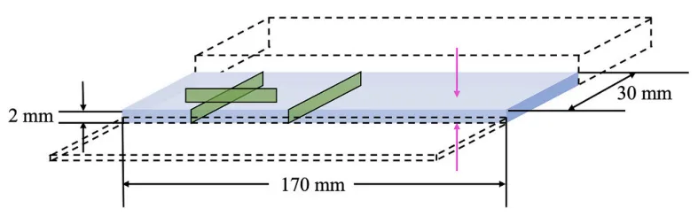
Fig. 2. Graphical representation of an as-cast part of 2 mm step thickness, with green boxes indicating the locations used for analysis using optical microscopy. The pink arrows indicate the two casting surfaces on which the skin layer is supposedly formed. (Adapted with permission from Dalai et al. [19]). (For interpretation of the references to color in this figure legend, the reader is referred to the web version of this article.)
Fig. 2. Graphical representation of an as-cast part of 2 mm step thickness, with green boxes indicating the locations used for analysis using optical microscopy. The pink arrows indicate the two casting surfaces on which the skin layer is supposedly formed. (Adapted with permission from Dalai et al. [19]). (For interpretation of the references to color in this figure legend, the reader is referred to the web version of this article.)